

3D View into the Structure of the Swine Flu (H1N1) Virus
Swine flu is a respiratory disease caused by a relatively new strain of the influenza virus known as H1N1.
It is a contagious viral infection transmitted by inhalation or ingestion of droplets containing virus from people sneezing or coughing.
These extremely small spherical viruses are only visible under an electron microscope.
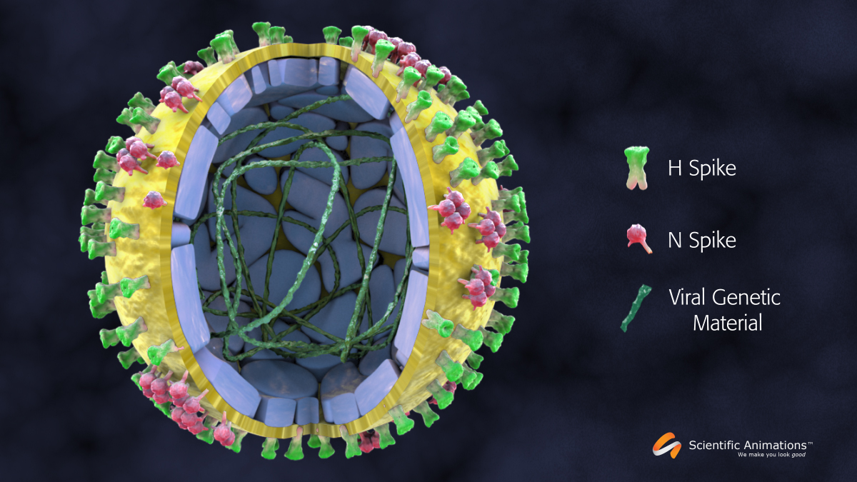
Here we give you a 3D view of this sub-microscopic nanoparticle, which is about 1000 times smaller than the width of a human hair along with details of its structure.
The core of the virus is genetic material that carries a blueprint for building copies of the virus once it enters the cell nucleus.
This genetic content is safeguarded by a hard protein shell, which enables it to safely travel from one human to another.
It is further wrapped in a viral membrane that is responsible for allowing the virus to infect healthy cells.
This viral membrane then has 2 types of projections on it –
- H Spikes or Hemagglutinins are responsible for attaching the virus to the cell receptor allowing it an entry into the cell.
- N Spikes or Neuraminidase help the newly formed viruses to disengage from the cell membrane enabling them to float freely in the system to infect other cells.


